Any Questions? Any Inquiries?
We are here to talk to you!
+94 -11 - 2697669
Tomey - EM 4000.

Specular Microscope
The EM-4000 is a specular microscope with a wide measurement range and auto analysis. Multi display functions and quick analysis enables smooth endothelium testing. Also database functions are installed to compare the observation of the patient pre and post surgery, or to re-analyze in several analysis methods.
Wide measurement range and image capture of surrounding areas
Using unique technology, a wide endothelium area of 0.25×0.54 mm can be viewed.
The EM-4000 can measure a total of 13 points including the center point, 6 peripheral positions and 6 parafovea positions, which provide more choices for imaging the endothelium area through corneal clouding.
Auto analysis and multi display functions
With built-in analysis software, 8 types of analysis values are automatically displayed. The layout of analysis results is selectable.
Captured images can be displayed in 4 ways (Photo / Trace / Area / Apex) making it clearer to see the endothelium
Dark area analysis function
More comfortable image capturing with higher speed
Display the analysis results 4 seconds (2 seconds/ eye) approx. after measurement. Touch alignment is simple, and smooth and speedy image capturing facilitates comfortable testing.
Internal database installed
A database is installed in the main unit. By displaying two data of one patient, it is possible to compare pre and post surgery. We also provide a patient list screen for patient identification.
Specifications
| Observation and analysis of corneal endothelium | |
|---|---|
| Measurement method | Non-contact |
| Photographing range | 0.25(W)×0.54(H)mm |
| Measurement mode | Auto / Manual / Auto alignment with 3D eye tracking method |
| Capturing position | Center + 12 peripheral points |
| Cornea thickness measurement | ✔ |
| Analysis method | Automatic analysis / L-count / Trace / Core Method |
| Analysis values | Number (the number of analyzed cells) CD (cell density) AVG (average cell area) SD (standard deviation of cell area) CV (coefficient of variation of cell area) MAX (maximum cell area) MIN (minimum cell area) 6A (hexagonal cell appearance rate) |
| Histogram | Area (Polymegathism: Distribution by areas) Apex (Pleomorphism: Distribution by polygonal shapes types) |
| Body | |
|---|---|
| Display | 10.4 inches, color LCD Touch panel |
| Dimensions and weight | 309(W)×491(D)×450(H) mm / approx. 22 kg |
| Power source | |
|---|---|
| Input voltage / frequency / power consumption |
100-240 VAC, 50/60 Hz, 100 VA |
Other Products
-
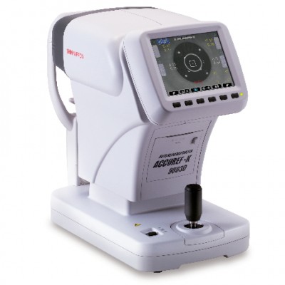
Rexxam (Shin-Nippon) - ACCUREF-K 9003D.
ACCUREF-K 9003D NEXT GENERATION HYBRID CONCEPT AUTO REFKERATOMETER
-
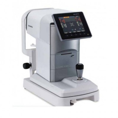
Rexxam (Shin-Nippon) - ACCUREF K-900/R-800.
ACCUREF K-900/R-800 AUTO REFRACTO METER
-

Ortopad - OCCLUSION EYE PATCHES.
OCCLUSION EYE PATCHES
-

Neotech - DS-2000 PLUS MANUAL SURGEON'S CHAIR.
MANUAL SURGEON’S CHAIR